
Date: 18th September 2015, Friday; Time: 10:30 – 16:00 hrs
Venue: MB 1017, First Floor, Minerva Building, Brayford Pool Campus, University of Lincoln, Lincoln LN6 7TS
A one-day research workshop on the theme “Building Models; Understanding Brain” was organised at the University of Lincoln, sponsored by the Royal Academy of Engineering (RAE). The workshop was organised by Dr. Piotr Suffczynski, who was visiting the School of Engineering for a duration of 1 month as a Distinguished Visiting Fellow awarded by the RAE, and Dr. Basabdatta Bhattacharya, who has been the Principal Investigator in the award.
Among the audience were Dr. Jun Chen, Sr. Lecturer, Ms. Elham Zareian, PhD student, from the School of Engineering; Dr. Simon Durrant, Sr. Lecturer, Dr. Louise O’Hare, Lecturer, from the School of Psychology, University of Lincoln.
PROGRAMME FOR THE DAY |
|
|
10.50 – 11.00 |
Welcome Basabdatta Sen Bhattacharya |
|
11.00 – 11.30 |
Mechanisms underlying different focal seizure onset patterns Yujiang Wang
|
|
11.30 – 12.00 |
Predicting neurosurgical outcomes in focal epilepsy patients using computational modelling Peter Taylor |
|
12.00 – 12.30 |
A multi-level view of deep brain stimulation induced effects on neurons and networks Nada Yousif |
|
12.30 – 13.45 |
Lunch break |
|
13.45 – 14.15 |
Revealing A Brain Network Endophenotype In Families WithIdiopathic Generalised Epilepsy Wessel Woldman |
|
14.15 – 14.45 |
Anti-NMDAR encephalitis causes changes in synaptic transmission dynamics in paediatric patients Richard Rosch |
|
14.45 – 15.00 |
Break |
|
15.00 – 15.30 |
Studying the role of thalamic interneurons with computational models Basabdatta Sen Bhattacharya |
|
15.30 – 16.00 |
On the origin of the harmonic frequencies in SSVEP signals Piotr Suffczynski |
Abstracts of the talks
Mechanisms underlying different focal seizure onset patterns
Yujiang Wang, University of Newcastle
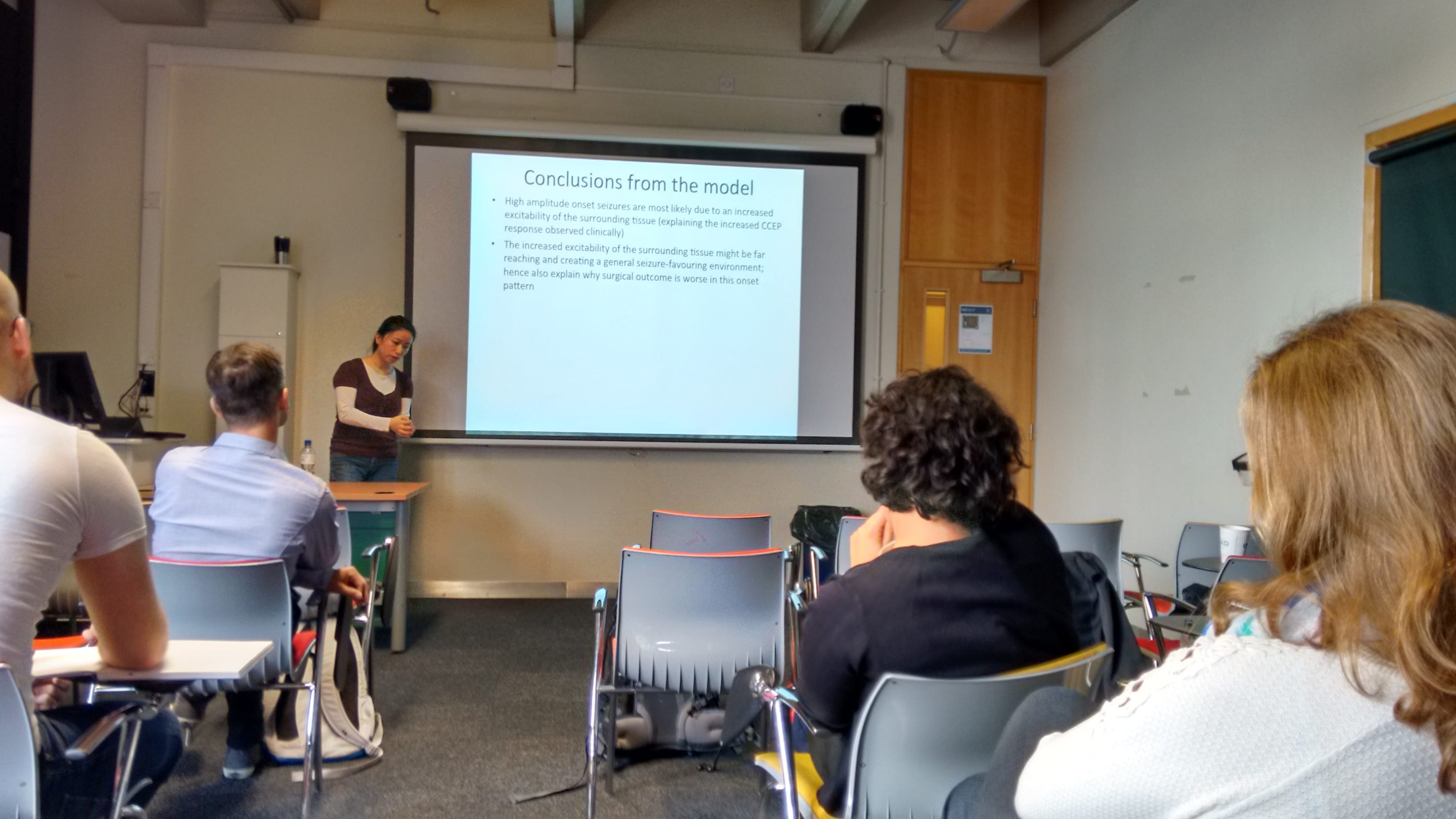
With the rise of new recording techniques in the last decade, clinical, experimental, and theoretical works have started to uncover a new understanding of neocortical focal seizure onset [1]. However, a long-standing observation has not been discussed in the context of the new discoveries yet: neocortical focal seizures typically begin with one of two typical waveform patterns — low amplitude fast oscillations (LAF), or high amplitude spikes/sharp waves (HAS) [2]. Interestingly, only the former of the patterns is associated with a good surgical outcome [3].
Based on previous work [1] we replicate these different onset patterns of intracranial recordings in a spatio-temporal computational model of a neocortical patch of tissue, and show that they are associated with different spatio-temporal patterns at the finer mesoscopic scale. The LAF is generated by initially independent patches of localized activity, which slowly evolve to a network of epileptic activity over time. In contrast, the HAS occurs as a transition to a globally bistable seizure state triggered by a local event. This indicates a difference in the spatial extent of the underlying pathology, which could explain the different surgical outcome associated with these two seizure onset patterns. Based on the results, we propose alternative treatment strategies for patients with the HAS onset pattern.
[1] Wang Y, Goodfellow M, Taylor P.N, and Baier G (2014) Dynamic mechanisms of neocortical focal seizure onset, PLoS CB, e1003787
[2] Perucca, P, Dubeau F, and Gotman J (2014) Intracranial Electroencephalographic Seizure-Onset Patterns: Effect of Underlying Pathology, Brain 137 (1): 183–96
[3] Lee S-A, Spencer D.D, and Spencer S.S (2000) Intracranial EEG Seizure-Onset Patterns in Neocortical Epilepsy, Epilepsia 41 (3): 297–307
A multi-level view of deep brain stimulation induced effects on neurons and networks
Nada Yousif, Imperial College London

Deep brain stimulation (DBS) is a clinical therapy, involving the surgical implantation of electrodes into disorder specific nuclei, used to treat the symptoms of movement disorders such as Parkinson’s disease and essential tremor (ET). The mechanisms through which the electrical stimulation leads to the observed clinical improvements remain unclear, to the extent that it is unknown how stimulation changes neuronal excitation. If we better understood these mechanisms, we could optimise the parameters setting process and minimise unwanted side effects. To study the effect of stimulation on the firing activity of axons, single neurons, and networks of neurons, I take a multi-level computational modelling approach. First I create a finite element model to calculate the distribution of electric potential induced by DBS and then applied the potential as an extracellular stimulus to multicompartment axon or neuron models. I have previously shown that such models can reveal detailed spatial effects of stimulation in the vicinity of the electrode. To study network level changes, I model the network proposed to be involved in generating pathological synchronous activity via a Wilson-Cowan approach. The parameter space of such a model can be explored to uncover regions which produced oscillatory thalamic activity in the typical ET frequency range (4-12Hz). Taken together, these models demonstrate a method of quantitatively assessing neuronal changes induced by DBS, to maximise therapeutic benefit and minimise unwanted side effects.
Predicting neurosurgical outcomes in focal epilepsy patients using computational modelling
Peter Taylor, University of Newcastle
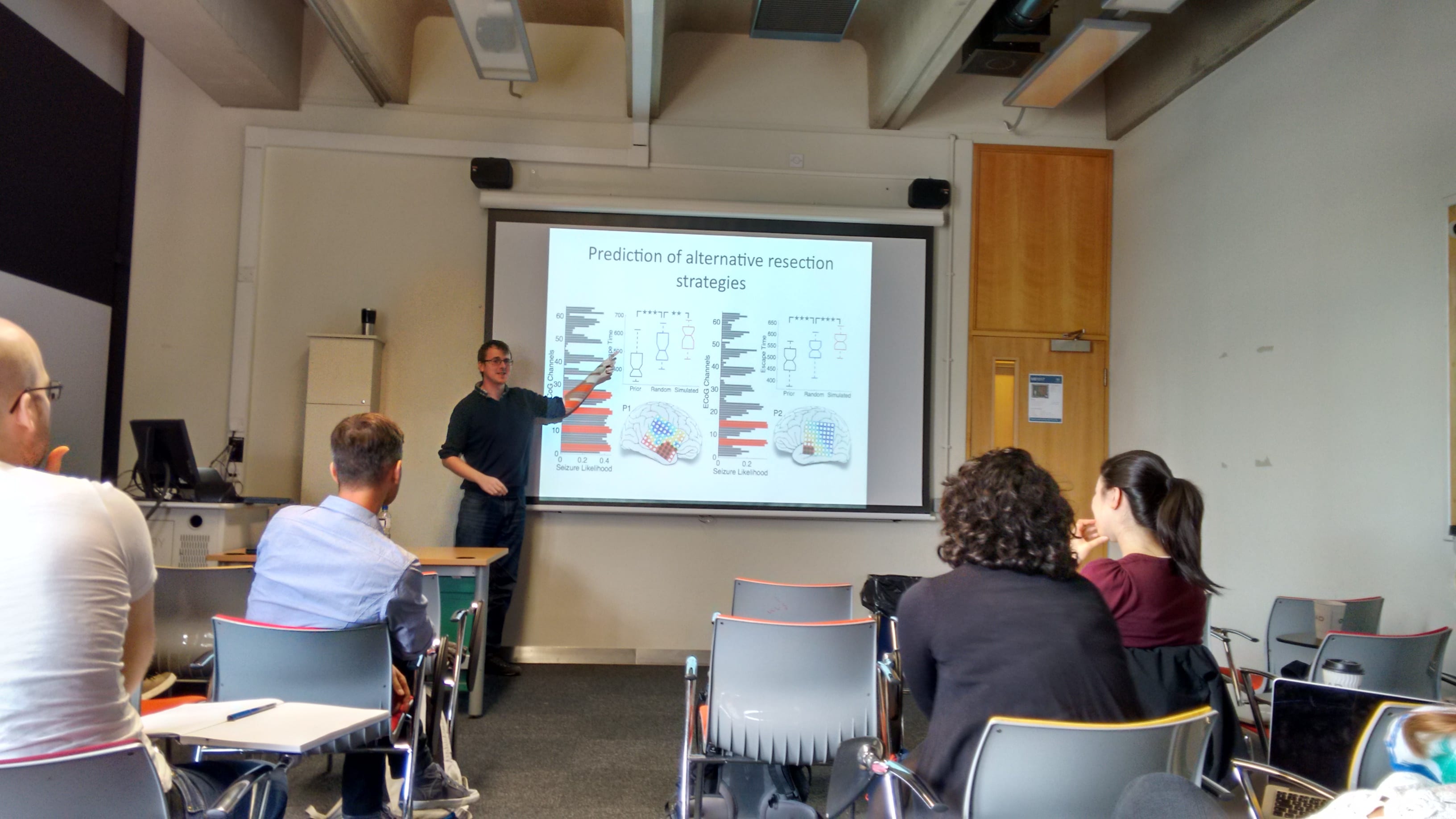
A third of patients with epilepsy are refractory to anti-epileptic drug treatment. For some of these patients with focal epilepsy, better seizure control can be achieved by surgical treatment in which the seizure focus is localized and resected while avoiding crucial cortical tissues. However surgical outcomes are unpredictable and many patients continue to have seizures, even after surgery. We propose a dynamical computational model in which model parameters are constrained by patient-specific data to predict surgical outcomes. We find that the model predicion accuracy is high (78%) and suggest this may be used as a tool to aid clinicians in presurgical evaluation of focal seizure patients.
Revealing A Brain Network Endophenotype In Families With Idiopathic Generalised Epilepsy
Wessel Woldman, University of Exeter
In [1] we studied resting state electroencephalogram (EEG) recordings from people with idiopathic generalised epilepsy (IGE) and their unaffected first-degree relatives, to test the assumption that abnormal brain network properties would manifest as a familial endophenotype. The study included 117 participants: 35 people with IGE, 42 unaffected first-degree relatives, and 40 normal controls, using scalp EEG. Network topology was derived using phase-locking factors, and significant differences were found in the 6-9-Hz band: degree distribution variance was greater in patients and relatives (p=0.0005, p=0.0009), mean degree was greater in patients and healthy controls (p=0.0064), and clustering coefficient was higher in patients and relatives than controls (p=0.0025, p=0.0013).
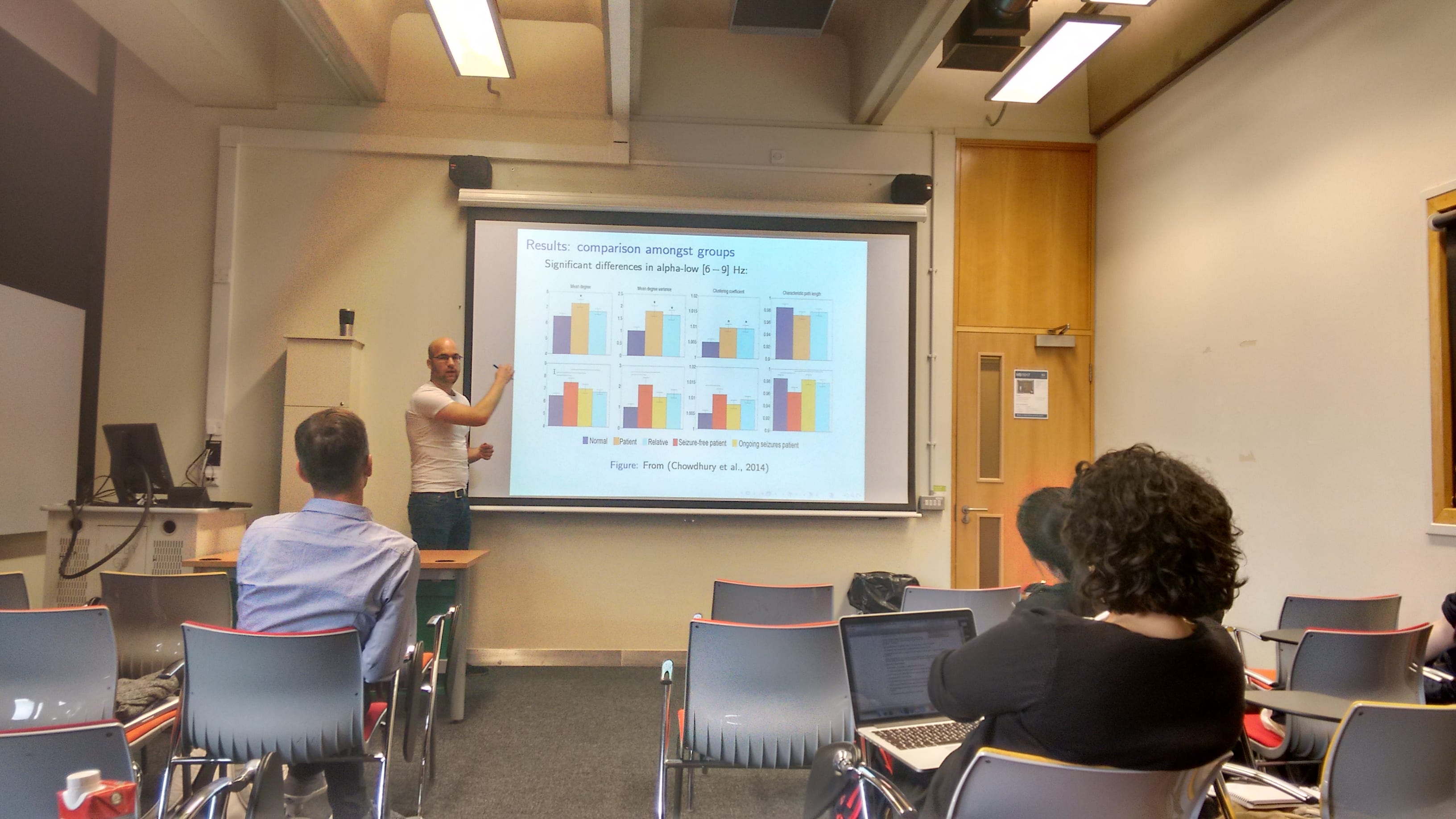
These findings suggest brain network topology differs between patients with IGE and normal controls, and that some of these network properties present themselves in unaffected relatives who do not themselves have epilepsy. This suggests brain network topology may be an inherited endophenotype of IGE, a necessary, although not a sufficient condition for epilepsy. Our findings support the concept that in order to understand how epileptic brains respond to clinical interventions it is essential to combine the study of complex brain networks with mathematical models that describe the dynamics of localised brain tissue.
[1] Chowdhury, F.A., Woldman, W., FitzGerald, T.H.B, Elwes, R.D.C, Nashef, L., Terry, J.R., Richardson, M.R. (2014) Revealing a Brain Network Endophenotype in Families with Idiopathic Generalised Epilepsy, PLoS ONE, vol. 9, no. 10, e110136
Anti-NMDAR encephalitis causes changes in synaptic transmission dynamics in paediatric patients.
Richard Rosch, University College London
Epileptic seizures are stereotyped paroxysmal neuronal events that can occur clinically in response to a wide variety of pathophysiological mechanisms. They are a common feature in the encephalitides, acute inflammatory conditions commonly caused by infectious, or autoimmune causes. In recent years, several discrete autoimmune encephalitis syndromes have been delineated, some of which are characterized by autoantibodies to cell-surface molecular targets with direct relevance for synaptic and neuronal function (Irani et al 2014, Ann Neurol 76). Yet there is only limited direct evidence linking the molecular pathophysiology with observable paroxysmal abnormalities in cortical neural dynamics in patients.
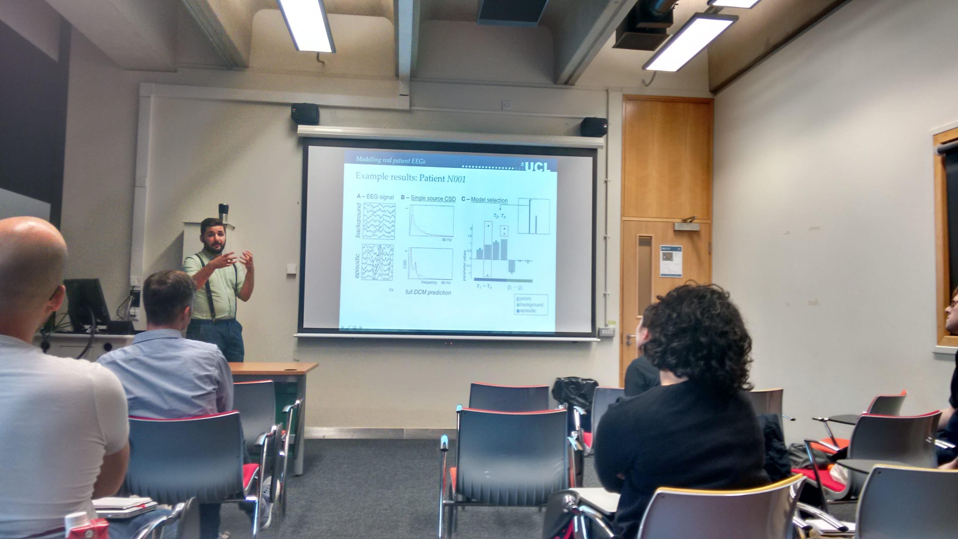
In this study, we investigate electroencephalographic (EEG) paroxysms in 8 paediatric patients with serologically proven Anti-N-methyl-D-aspartate receptor (Anti-NMDAR) associated encephalitis. Using dynamic causal modelling, we estimate parameters of a biologically plausible neural network model, empirically based on the EEG observations (Friston 2014, Brain 137). From the model, the changes in intrinsic connectivity among its constituent inhibitory and excitatory neuronal subpopulations are estimated for different time windows of around the paroxysmal events.
The results of this analysis allow empirical comparison of the likelihood of different mechanistic models explaining the observed EEG paroxysms. Despite phenomenological diversity in the EEG paroxysm observed in the patients reported here, in each individual patient they were best explained by changes in synaptic transmission time constants, without changes in synaptic connection strengths.
This study is a first step in (i) validating empirically informed models of EEG paroxysms; and (ii) identifying the contributions of molecular pathophysiology to observable pathological neuronal responses noninvasively. With an increasing number of candidate molecular causes for epileptic paroxysms (including genetic mutations and autoantibodies), the approach presented here may prove valuable in testing potentially treatment-relevant hypotheses about the network mechanisms through which molecular abnormalities are manifest as a clinical phenotype.
Studying the role of thalamic interneurons with computational models
Basabdatta Sen Bhattacharya, University of Lincoln
Literature review suggests that the role of thalamic interneurons (IN) are often ignored in thalamocortical models, the focus being on the connectivity between the thalamocortical relay (TCR) cells and the cells of the thalamic reticular nucleus (TRN). In this talk, I will present some recent research results that show the drastic effect that the IN has on the TRN and TCR population behaviour in a simple population model. This was when the model input was: (a) Gaussian noise and validated with EEG of the state of quiet wakefulness; (b) pulse inputs at frequencies of 6, 8 and 10 Hz and validated respectively with SSVEP collected under application of flickering stimulus at these frequencies. Similar study using spiking neural networks modelling thalamic cell populations and implemented on the SpiNNaker machine also shows the significant effect of IN on the TCR and TRN behaviour when all cells are operating in the tonic mode. This is however not the case when the TRN is operating in the bursting mode. I will discuss the implications of these observations during the talk.
On the origin of the harmonic frequencies in SSVEP signals
Piotr Suffczynski, RAE Distinguished Visiting Fellow
University of Warsaw, Poland
The steady state visual evoked potentials (SSVEP) are oscillatory brain responses to periodic light stimulation, observable in EEG signals. Frequency of these responses corresponds to the stimulus frequency and its harmonics. Despite the SSVEP signals have widespread applications in cognitive and clinical neuroscience, and engineering, the mechanisms responsible for its generation are not fully understood. Since the very first reports on SSVEP in 1966, they have been commonly assumed to be stationary with power and amplitude stable over time. Using prolonged stimulation paradigm (60-seconds) long we show that instantaneous power of SSVEP may change significantly for the large majority of subjects. Another feature, which still awaits an explanation, is related to harmonic components in the response. In the present study we explore the origin of the
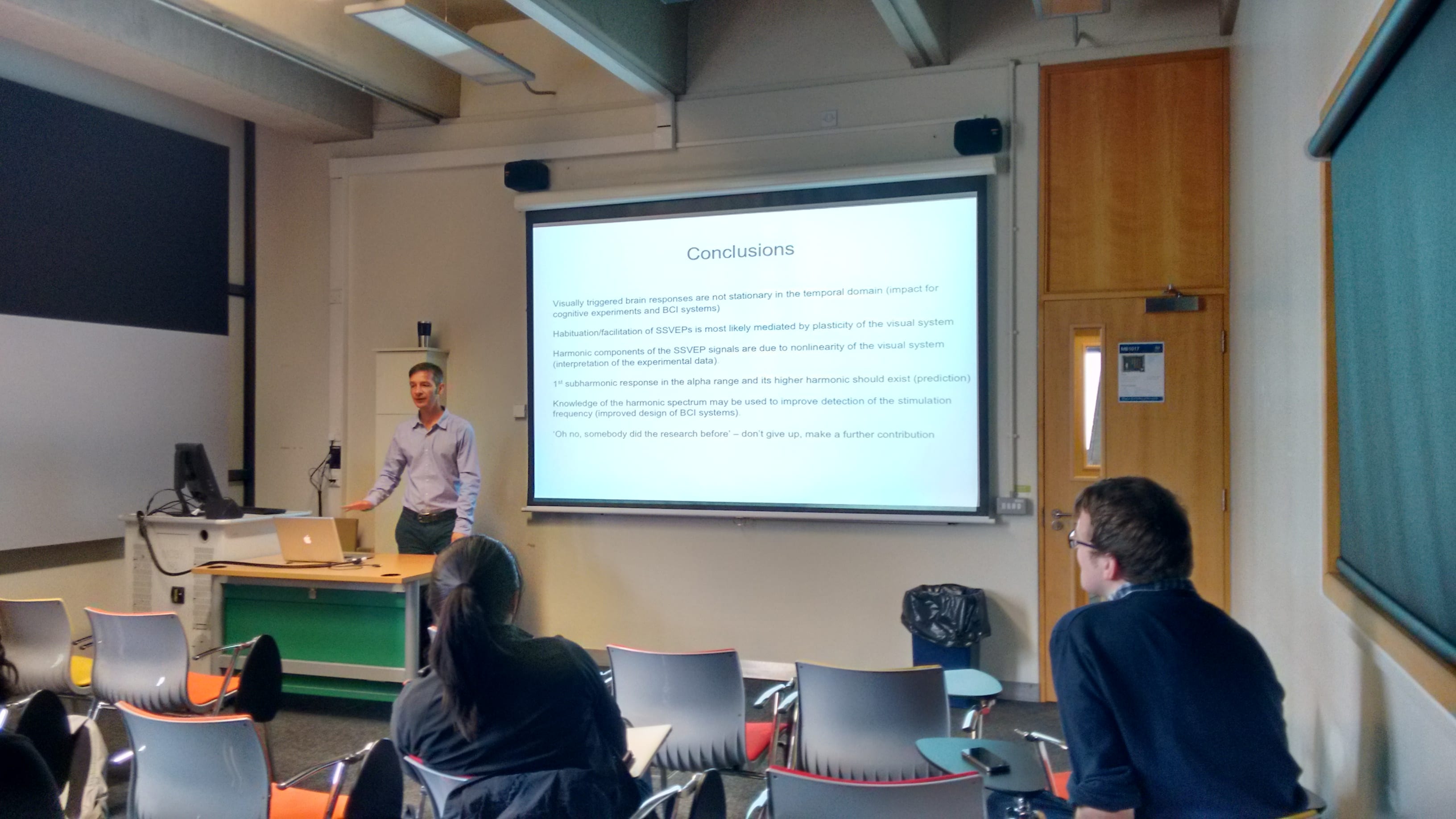
harmonic responses using computational neural mass model that takes into account the general properties of the cortical networks. The spectra of modeled SSVEP signals share many similarities with experimental observations what allows us to suggest the mechanisms responsible for their generation, identify key factors shaping response spectra and also generate new testable predictions.